Using High Performance Computing for Biomechanical Finite Element Models

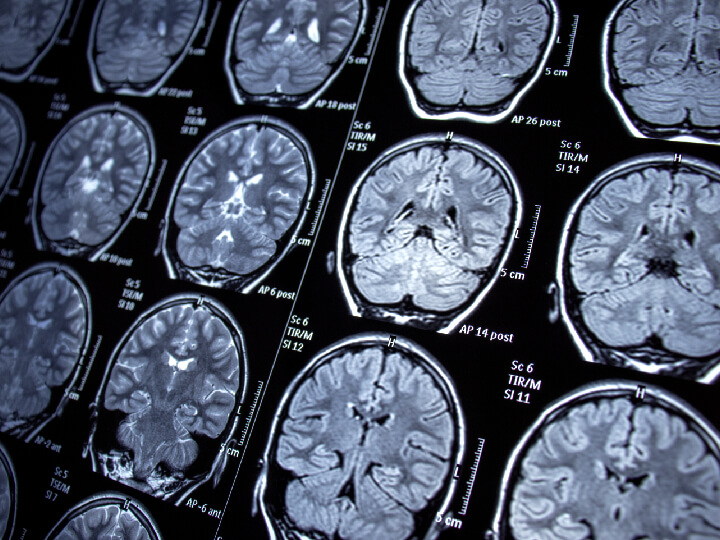
Brain-shift during neurosurgery can compromise the validity of preoperative images when identifying the locations of tumors and vital structures in neuronavigational platforms. Brain shift is a deformation of the brain that occurs during neurosurgery. It can be caused by several factors indirectly related to surgery such as gravity, head positioning, fluid drainage, use of drugs, changes in intracranial pressure or swelling of brain tissue. Although several studies have used various finite element models (FEMs) to successfully predict inward brain-shift, they are time-consuming and their CPU performance is not clinically compatible.
Research scientist Anne-Cecile Lesag has worked to develop a model to describe inward brain-shift with viscoelastic biomechanical modeling varying three surgery parameters: craniotomy position, cerebrospinal fluid drainage level and intraoperative head position. In this seminar, Lesag demonstrates that the combination of HPC generated FEM training data and a U-net approach is a promising solution for clinically compatible performance.
Throughout the seminar, Lesag discussed prior research that structured her project, such as literature indicating that problems with slowness in workflow of patient-specific FEM could be solved with deep learning. She detailed the deep learning workflow model her team followed, as well as further questions they developed throughout their studies.
“I hope that with deep learning, we can one day develop a system in which we can avoid having to build a finite element simulation,” said Lesag. “To not have to spend one or two days building a patient profile is the goal.”
Anne-Cecile Lesage is a research scientist at M D Anderson Cancer Center. She has 17 years of experience in the field of applied mathematics (numerical analysis, finite elements, finite difference, inverse methods) and applied physics (hydraulics, hydrodynamics, solid mechanics, geophysics). She joined M D Anderson in 2018, more specifically the Morfeus Lab supervised by Professor Kristy Brock in the Imaging Physics department. Her current research focuses on modeling soft tissue mechanics for medical applications. A part of the image-guided cancer therapy program, she is developing patient-specific finite element modeling workflows to predict tissue deformations for applications in brain and lung cancer surgery.